What Is Normal Myocardial Perfusion
Coronary artery blood flow can be assessed and the scans can also be used to accurately determine the left ventricular ejection fraction the end-systolic volume of the left ventricle regional wall motion and wall thickening 4. Its also known as myocardial perfusion imaging or a.

Normal Myocardial Perfusion Imaging Study In A 86 Y O Patient No Download Scientific Diagram
A low urine output and confusion are signs of decreased tissue perfusion.

What is normal myocardial perfusion. Myocardial perfusion imaging MPI is a form of functional cardiac imaging used for the diagnosis of ischemic heart diseaseThe underlying principle is that under conditions of stress diseased myocardium receives less blood flow than normal myocardium. 22 23 Myocardial hibernation has been demonstrated in infarcted areas of the myocardium and also in areas remote from the area of infarct but. Myocardial stunning occurs after acute myocardial infarction as evidenced by patients treated with reperfusion therapy who have shown near normal recovery by 2 weeks.
A myocardial perfusion scan uses a tiny amount of a radioactive substance called a radioactive tracer. Orthopnea is a sign of left-sided heart failure. Acute myocardial infarction.
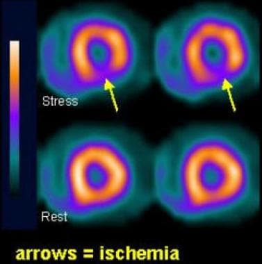
A mild decreased uptake in basal inferior wall and septum. Crackles edema and weight gain should be monitored closely but the levels are not as high a priority. On the scan the areas where tracer has been absorbed look different from the.
Normal myocardial perfusion SPECT MPS in a 55-year-old man referred because of positive exercise stress test. A cardiac specific radiopharmaceutical is administered eg 99m. The tracer travels through the bloodstream and healthy heart muscle absorbs it.
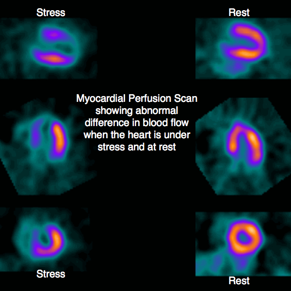
MPI is one of several types of cardiac stress test. Both stress and rest images demonstrate normal radiotracer distribution. Currently nuclear myocardial scans include both perfusion and gated wall motion images.
A cardiac perfusion test tells your doctor if the muscles of your heart are getting enough blood.

Myocardial Perfusion Images From Normal Stress Only Study Performed On Download Scientific Diagram

Figure Normal Myocardial Perfusion Scan Showing Location Of Segments Contributed By Ghufran Adnan Statpearls Ncbi Bookshelf
Myocardial Perfusion Spect Background Indications Contraindications

Myocardial Perfusion Images From Normal Stress Only Study Performed On Download Scientific Diagram
Normal Stress Myocardial Perfusion Scan In A 48 Year Old Lady Planned Download Scientific Diagram
2

Myocardial Perfusion Imaging Shows Promise In First Time Heart Failure
Myocardial Perfusion Scan Heart Of England Cardiology
![]()
Nuclear Medicine 3 Myocardial Perfusion Imaging Nursing Times
0 Response to "What Is Normal Myocardial Perfusion"
Post a Comment